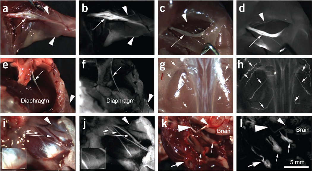
Figure 1.
Whole-body survey of nerves in mice (n = 3) 4 h after injection with 450 nmoles of FAM-NP41. (a,b) Brachial plexus. Reflectance image (a) showing left brachial plexus (arrow). Smaller branches (~50–100 µm) are easily seen (arrowheads) with fluorescence labeling (b) but not in reflectance image. (c,d) Sciatic nerve. Reflectance image (c) showing right sciatic nerve (arrow). Many more small branches (~50–100 µm) are seen (arrowheads) with fluorescence imaging (b) compared to reflectance. (e,f) Phrenic nerve. Reflectance image (e) showing left phrenic nerve (arrow) descending from the mediastinum to innervate the diaphragm. Note that the nerve is seen as a single linear fluorescent structure (arrow, f) compared to the bundle of nerve and connective tissue seen with reflectance (arrow, e). Arrowhead points to an intercostal nerve which is easily seen with fluorescence but not with reflectance. (g,h) Dorsal cutaneous nerves. Reflectance image (g) showing the dorsal musculature. Fluorescence imaging highlights the dorsal intercostal nerves (arrows, h) that are not easily seen with reflectance. (i,j) Facial nerve. Main facial nerve branches (arrows) are easily seen with both reflectance (i) and fluorescence (j). However, a small branch of nerve arborization (arrowhead) leading to the upper face can be distinguished from surrounding tissue with fluorescence but not with reflectance. Insert shows arborizations (~50 µm diameter) of the lower division of the facial nerve that can be easily seen with fluorescence labeling. (k,l) Dorsal view of skull base. Reflectance image (k) showing left facial nerve (large arrow) wrapping around the ear, trigeminal nerves (small arrows), optic nerves (large arrowhead) and optic chiasm (small arrowhead). Fluorescence image (l) shows fluorescence labeling of the facial and trigeminal nerves (peripheral nervous system) but not the optic nerves and chiasm (central nervous system). Scale bar (a–l), 5 mm; inserts (i and j), 1 mm.

