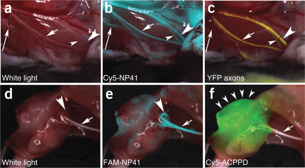
Figure 4.
NP41 can highlight buried nerve branches invisible by standard illumination. (a–c) Right facial nerve and its arborizations in a thy1-YFP mouse treated with Cy5-NP41, viewed by (a) white light reflectance, (b) Cy5 fluorescence (pseudocolored cyan) overlaid on reflectance and (c) YFP fluorescence (pseudocolored yellow), also overlaid on reflectance. The short arrow marks a nerve branch visible by all three imaging modes. The arrowheads point to branches that are difficult to differentiate from muscle fascia in reflectance, but clearly distinguishable in both fluorescence images. The long arrow indicates a deeply buried branch visible only by Cy5-NP41 due to the better penetration of far-red wavelengths. (d–f) Left sciatic nerve (arrow) and its arborization in a mouse with a syngeneic 8119 mammary tumor graft17,18, viewed by (d) white light reflectance, (e) FAM fluorescence 2 h after intravenous injection of NP41 (150 nmoles) (pseudocolored cyan, overlaid on reflectance) and (f) Cy5 fluorescence (pseudocolored green, overlaid on reflectance) from conjugates of activatable cell-penetrating peptides and dendrimers (ACPPDs). The large arrowheads in d and e point to a nerve branch buried under tumor, visible only by FAM fluorescence. Small arrowheads in f denote tumor. See Supplementary Video 1.

