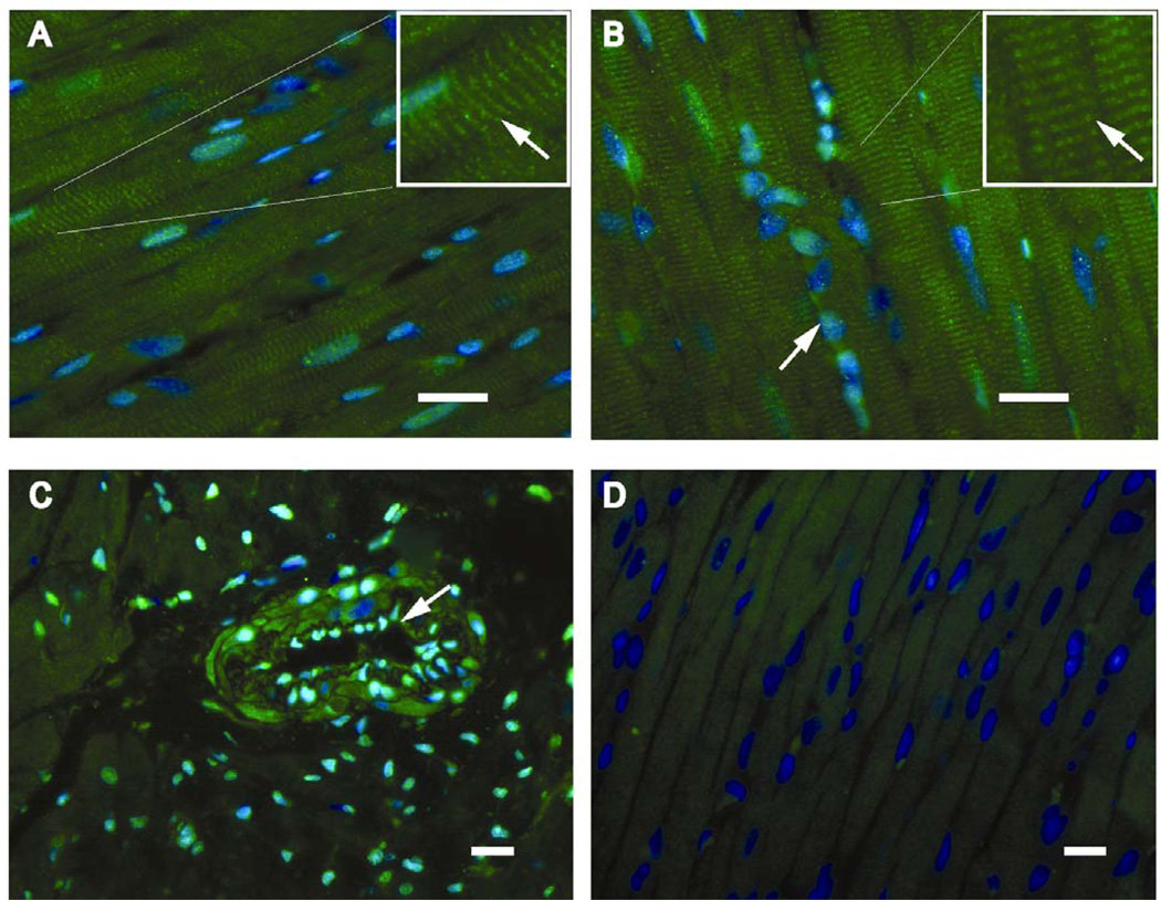
Figure 3. Distribution of XO/XDH cardiomyocytes in sham and 24 hr ACF left ventricles.
Immunohistohemistry demonstrates XO/XDH in cardiomyocytes in coordination with Z-lines (arrows), in sham (A) and 24 hour ACF LVs (B) the box in the top right of each panel is a higher magnification view of the same image. In addition, panel B demonstrates XO/XDH in interstitial cells (fibroblasts and endothelial cells) in which nuclei stained with DAPI (blue) take on a more green appearance. In C, cross section of an arteriole in a sham LV demonstrates XO/XDH staining in endothelial cells (arrow) and in smooth muscle cells. The immunoadsorbed antibody (D) demonstrates the blue staining of nuclei with DAPI and lack of XO/XDH staining supporting the specificity of the XO/XDH antibody. White bar represents 20 µm (note different magnifications).

