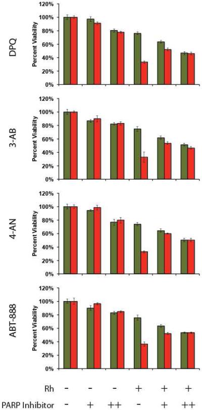
Figure 7.
PARP inhibition assay. HCT116N and HCT116O cells were plated in 96-well format at densities of 5 × 10⁴ cells/well and treated with 0 μM (“−”) or 20 μM (“+”) of [Rh(HDPA)2chrysi]3+ with or without PARP inhibitor (“-” = 0 μM DPQ, 3-AB, 4-AN, or ABT-888; “+” = 25 μM DPQ, 2 mM 3-AB, 10 μM 4-AN, or 5 μM ABT-888; “++” = 50 μM DPQ, 3 mM 3-AB, 20 μM 4-AN, or 10 μM ABT-888). After 72 hours, the cells were labeled with MTT for 4 hours. The resulting formazan crystals were dissolved by addition of 10% SDS acidified with 10 mM HCl, and absorbance was measured at 570 nm. All four PARP inhibitors confer protection from rhodium-induced MMR-dependent toxicity, reducing the differential activity significantly.

