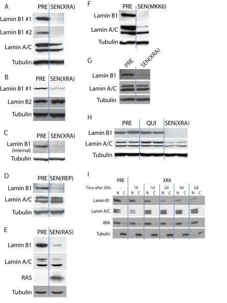
FIGURE 1:
Lamin B1 loss is associated with multiple types of cellular senescence. (A) Lamin B1 declines in DNA damage–induced senescence. HCA2 cells were mock irradiated (PRE) or irradiated (10-Gy x-rays) and allowed to senesce (SEN(XRA)). Whole-cell lysates were analyzed by Western blotting using either of two unrelated lamin B1 antibodies that recognize C-terminal epitopes. (B) Lamin B2 does not decline in DNA damage–induced senescence. HCA2 cells were mock irradiated (PRE) or irradiated and allowed to senesce (SEN(XRA)). Whole-cell lysates were analyzed by Western blotting. (C) Lamin B1 declines in SEN(XRA) cells. BJ cells were mock irradiated (PRE) or irradiated and allowed to senesce (SEN(XRA)). Whole-cell lysates were analyzed by Western blotting using a third lamin B1 antibody that recognizes an internal epitope. (D) Lamin B1 declines in replicative senescence. HCA2 cells were cultured until replicative senescence (SEN(REP); ∼70 population doublings). Whole-cell lysates were analyzed by Western blotting. (E) Lamin B1 declines in RAS-induced senescence. HCA2 cells were infected with a lentivirus lacking an insert (PRE) or expressing oncogenic RASV12 and allowed to senesce (SEN(RAS)). Whole-cell lysates were analyzed by Western blotting. (F) Lamin B1 declines in MKK6-induced senescence. HCA2 cells were infected with a lentivirus lacking an insert (PRE) or expressing a constitutively active MAP kinase kinase 6 mutant (MKK6EE) and allowed to senesce (SEN(MKK6). Whole-cell lysates were analyzed by Western blotting. (G) Lamin B1 declines in WI-38 cells after XRA. WI-38 cells were irradiated (SEN(XRA)) and allowed to senesce. PRE cells were mock irradiated. Whole-cell lysates were analyzed by Western blotting. (H) Lamin B1 does not decline in quiescent cells. HCA2 cells were cultured in media containing 10% serum (PRE), serum-free media for 48 h to induce quiescence (QUI), or irradiated and allowed to senesce (SEN(XRA)). Whole-cell lysates were analyzed by Western blotting. (I) Lamin B1 declines within 48 h after induction of senescence by irradiation. HCA2 cells were mock irradiated (PRE) or irradiated (XRA). Nuclear (N) and cytoplasmic (C) extracts collected at the indicated time points were analyzed by Western blotting. RPA serves as a loading control for the nuclear fraction; tubulin serves as a loading control for the cytoplasmic fraction.

