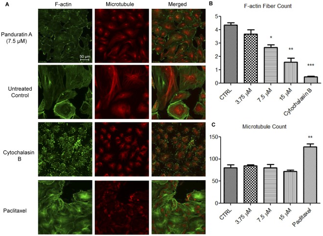
Figure 8. Effects of PA on cytoskeletals system of HUVECs.
(A) HUVECs were treated with PA, paclitaxel, cytochalasin B, or medium alone (untreated control) for 16 h. HUVECs were fixed and stained with DY554-phalloidin for F-actin, and anti-tubulin antibody for microtubules, respectively. Images were acquired on Cellomics ArrayScan HCS Reader. Number of (B) F-actin (stress fiber), and (C) Microtubule fibers count were analyzed by Cellomics Morphology BioApplication. Data are expressed as means ± SEM of three independent experiments. Statistical significance is expressed as ***, P<0.001; **, P<0.01; *, P<0.05 versus untreated control. Scale bar indicates 50 µm.

