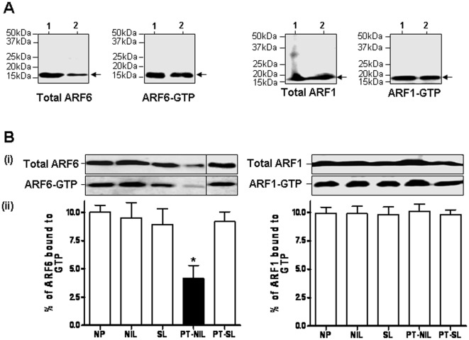
Figure 5. The GST-pulldown analysis of active ARF1 and ARF6 levels in the myometrium.
A. Analysis of the ARF1-GTP and ARF6-GTP levels in human placenta and myometrium. Lanes 1, Placenta and 2, NP myometrium. B. The GST-pulldown analysis of active ARF1 and ARF6 levels in human myometrium of NP, NIL, SL, NP-SL and PT-NIL. The tissue lysates from NP, NIL, SL, PT-NIL and PT-SL were incubated with GST-GGA3 PBD resin to precipitate ARF1-GTP or ARF6-GTP as described in the materials and method section. The total ARF1 and ARF6 expression in the cell lysates (upper panel) and the activated ARF1 and ARF6 that was pulled down with GST-GGA3 PBD (lower panel) were detected by immunoblotting using an anti-ARF1 rabbit polyclonal and an anti-ARF6 mouse monoclonal antibody respectively (i). The intensity of the bands was quantified by densitometric scanning. The data were expressed as percentage of total ARF1 or ARF6 that is GTP bound. The histogram represents an average of 4 samples for each condition (ii).

