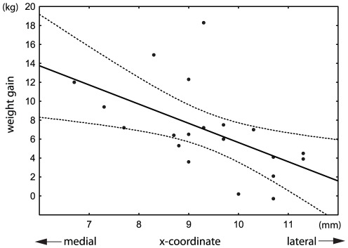
Figure 3. Weight gain in 20 patients with Parkinson's disease in relation to the mediolateral position of the active contact with bilateral STN-DBS (r = −0.55, p<0.01).
Only one active contact (more medial contact from both hemispheres) was used in each patient. The x-coordinate represents the distance of the active contact from the wall of the third ventricle. Each millimeter in the medial direction was associated on average with a 1.6-kg increase in body weight. Dotted lines denote the 95% confidence interval of the regression line.

