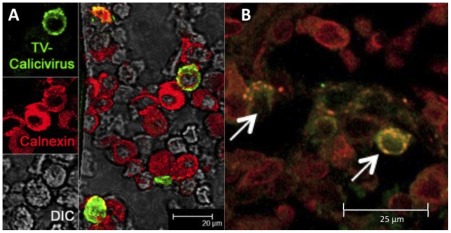
Figure 5. Colocalization of TV and calnexin antigens inside the HC55 macaque's duodenum lamina propria.
A– While most of the cells are calnexin-single-positive, simultaneous presence of both TV (green) and calnexin (red) antigens is seen in some of the (yellowish) cells. DIC– differential interference contrast. B– Spectral overlap of TV and calnexin antigens is shown by arrows.

