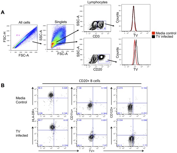
Figure 6. Flow cytometry detection of TV antigens-containing cells in vitro.
A– PBMCs isolated from healthy rhesus macaques were used for in vitro inoculation with TV. After being cultured in vitro for 6 days, cells were sorted out into CD3+ T cell and CD20+ B cell populations. Presence of TV antigens was detected in CD20+ B cells as shown by histograms (black peak). B– The CD20+ B cells were further subdivided into populations expressing the HLA-DR, CD11c or CD123 antigens. Presence of TV was revealed predominantly in CD20+HLA-DR+ cells while some of the other lymphocyte populations including the CD20+CD11c+ and CD20+CD123+ B cells also contained TV antigens.

