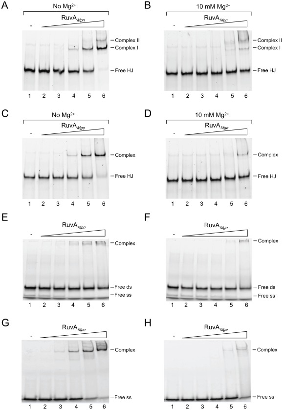
Figure 2. Binding of RuvAMpn and RuvAMge to HJs and other oligonucleotide substrates.
(A) Binding of RuvAMpn to HJ substrate HJ 1.1 in the absence of Mg2+. The DNA-binding reactions were performed as indicated in Materials and Methods. Reactions were performed in volumes of 10 μl and contained 12.3 nM DNA substrate and either 0 nM (marked ‘-’, lane 1), 27 nM (lane 2), 81 nM (lane 3), 243 nM (lane 4), 729 nM (lane 5) or 2.2 μM (lane 6) of RuvAMpn. Reaction products were electrophoresed through 8% polyacrylamide gels and analyzed by fluorometry. The positions of unbound HJ 1.1 (Free HJ) and RuvAMpn/HJ complexes (Complex I and II) are indicated at the right-hand side of the gel. (B) Binding of RuvAMpn to HJ substrate HJ 1.1 in the presence of 10 mM Mg2+. Reactions were carried out in a similar fashion as in (A). (C, D). Binding of RuvAMge to HJ substrate HJ 1.1 in the absence (C) or presence of 10 mM Mg2+ (D). (E, F) Binding of RuvAMpn (E) and RuvAMge (F) to double-stranded (ds) oligonucleotide HJ11/HJ11rv. The positions of the ds substrate (Free ds) and residual non-annealed oligonucleotide HJ11 (Free ss) is indicated at the right-hand side of the gels. (G, H) Binding of RuvAMpn (G) and RuvAMge (H) to single-stranded (ss) oligonucleotide HJ11. The reactions shown in panels (C) to (H) were carried out similarly as in (A).

