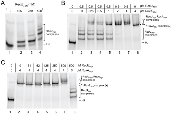
Figure 4. The interaction between RuvAMge and RecUMge on HJs.
(A) HJ-binding by RecUMge. The DNA-binding reactions were performed in a similar fashion as described in Fig. 3. Reactions were performed in volumes of 10 μl and contained 12.3 nM HJ 1.1 and the indicated concentrations of RecUMge. The positions of unbound HJs (HJ) and RecUMge-HJ complexes are depicted at the right-hand side of the gel. (B) The binding of RuvAMge to RecUMge-HJ complexes. RecUMge (0.5 μM) was incubated with HJ 1.1, followed by the addition of RuvAMge (at different concentrations, as indicated above the lanes). The nature of the various protein-DNA complexes is indicated at the right-hand side of the gel; RuvAMge-HJ complexes are indicated with a dot (•). (C) The binding of RecUMge to RuvAMge-HJ complexes. RuvAMge (4 μM) was incubated with HJ 1.1, followed by the addition of RecUMge (at various concentrations, as indicated above the lanes). The labeling of the figure is similar to that shown in (B).

