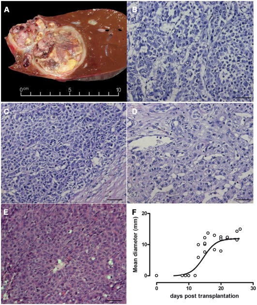
Figure 1. Histological appearance of the primary tumour and xenografts.
Cross section of the explanted liver revealed multifocal lesions with a heterogeneous encapsulated tumour (A). Three areas of the primary liver tumour show epithelial and carcinoma-like cell morphology (B, C, D). Tumours generated in mice had a high cellular density (E). Tumour passages in mice led to tumour nodules growing exponentially in the second week after xenotransplantation, reaching a mean diameter of 12 mm within 10 days (F). Bars represent 50 µm.

