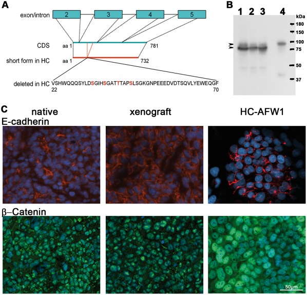
Figure 3. Expression of β-catenin and E-Cadherin in HC-AFW1 cells.
(A) β-Catenin in HC-AFW1 cells revealed a deletion of a polypeptide from amino acids (aa) 22 to 70 coded on exon 3. In this region 3 serine residues and a threonine amino acid are depicted in red; as phosphorylation sites for GSK3. (B) Western blot analysis revealed a main short form of β-catenin in cultured HC-AFW1 cells (1), in xenografts (2) and in the primary tumour (3). A larger form was found in the native liver (4)(arrowheads). (C) E-cadherin in native and xenograft tissue and in cultured HC-AFW1 cells was located at cell-cell contact (red fluorescence). All specimen tumour cells showed green fluorescence staining for β-catenin in the cell nucleus, and in some cells a membrane localization. Cultured HC-AFW1 cells had notably stronger β-catenin staining in the nuclei. DAPI counterstaining indicates the cell nuclei (blue fluorescence).

