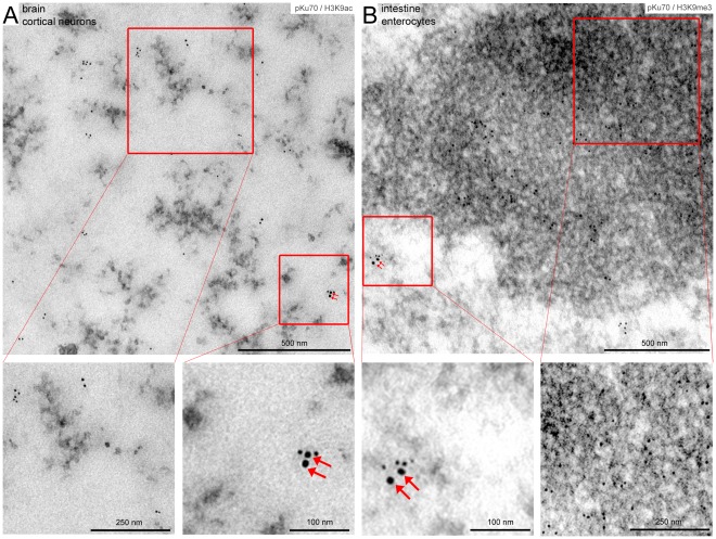
Figure 3. Co-labeling of pKu70 with euchromatic (H3K9ac) and heterochromatic marks (H3K9me3) analyzed 40 min after irradiation with 6Gy.
TEM micrographs of double-labeling of pKu70 (10-nm beads) with H3K9ac or H3K9me3, respectively, (6-nm beads) at different magnifications. Immunogold particles directed against H3K9ac are sparsely scattered within electron-lucent euchromatin (A), while gold-particles directed against H3K9me3 are highly enriched in electron-dense heterochromatin (B). Radiation-induced pKu70 clusters co-localize with H3K9ac (A) and H3K9me3 (B), suggesting that pKu70 allows the detection of euchromatic and heterochromatic DSBs.

