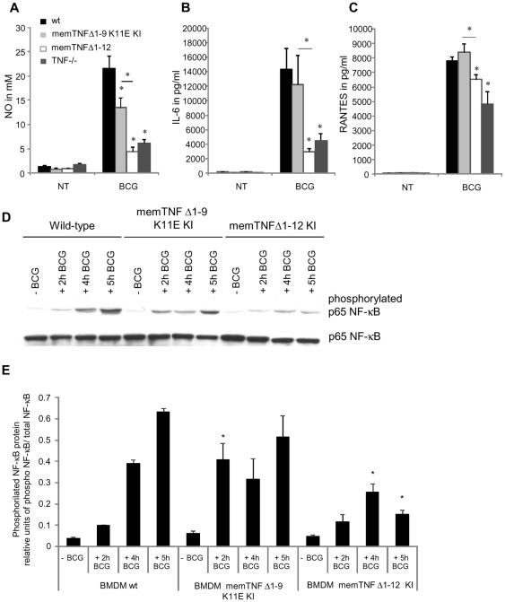
Figure 8. BMDM nitric oxide, IL-6, RANTES production and p65 NF-κB activation by M. bovis BCG infection.
NO (A), IL-6 (B) and RANTES (C) levels were assessed in supernatant of BMDM 24 hours after M. bovis BCG infection. These results are representative of two independent experiments and values are represented as mean ± SEM (n = 3 animals per group, assayed in triplicate) *p<0.05. (D) Phosphorylated and unphosphorylated p65 NF-κB proteins were detected by western blot in BMDM not infected or infected with M. bovis BCG at different time points. (E) Quantification of phosphorylated p65 NF-κB on western blot was done by Quantity One® analysis software on two different experiments. Results are expressed as mean ± SEM of relative units of phosphorylated p65 NF-κB/total p65 NF-κB (n = 2/group). *, p<0.05 compared to wild-type BMDM.

