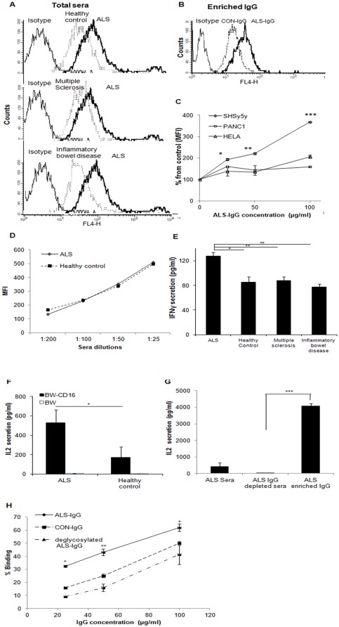
Figure 4. Coupling of IgG to target neuroblastoma and NSC34 cells and to CD16 on effector cells.
FACS histograms presenting a shift in binding of serum pools of ALS patients to human neuroblastoma cells in comparison to binding of healthy control (CON), inflammatory bowel disease and multiple sclerosis serum pools to neuroblastoma cells (A); FACS histogram presenting shift in binding of purified IgG from serum pools of ALS patients to neuroblastoma cells relative to binding of purified IgG from healthy control (CON) (B); Dose-dependent coupling of purified ALS-IgG to human PANC1, HeLa, and neuroblastoma cells performed as described in A(C); Mean fluorescent intensity (MFI) calculated relative to control sample containing cells and serum that was free of IgG. Dose-dependent coupling of ALS-IgG to mouse NSC34 cells was performed as described above (D); Secretion of IFNγ by enriched human peripheral NK cells in response to interactions with pools of ALS, inflammatory bowel disease patients, patients of multiple sclerosis, and healthy control (CON) sera (E); Secretion of IL-2 by BW-CD16 transfectants or BW cells in response to interactions with pools of ALS and healthy control sera (F), and in response to interactions with ALS-IgG and ALS IgG-depleted sera (G). Comparing the specificity of dose-dependent coupling of PNGase F-treated or untreated IgG of ALS patients and of the IgG of healthy volunteers, to CD16 (H). Data represent the mean ± SD of triplicate measurements from independent duplicate experiments. Pools of healthy and patient samples contained a mixture of four individual serum samples with similar glycan amounts represented in peaks 12 and 13. Statistical significance, *** p<0.005, ** p<0.01 and * p<0.05, versus the appropriate controls in each panel.

