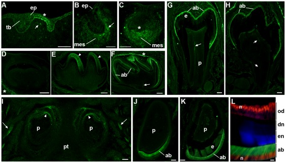
Figure 1. The expression of α7GFP changes during different developmental stages of tooth development.
A) Sagittal sections prepared of the E11.5 oral cavity shows α7GFP expression, as detected by immunohistochemical staining for GFP expression, in the thickened dental lamina (asterisk) of the oral epithelium (ep) adjacent to or between developing tooth buds (tb). Only occasionally do cells of the underlying tissues including developing mesenchyme (mes) express detectable α7GFP (arrow). B) E13.5 epithelial α7GFP is no longer detected, but some cells in the developing mesenchyme (mes) at the border with the invaginated epithelium express α7GFP (arrow head). C) E14.5 (bud stage) cellular expression of α7GFP is enriched in cells of the condensing mesenchyme where it is particularly intense adjacent to overlaid epithelium and the primary enamel knot (asterisk). D) The expression of α7GFP rapidly diminishes after E14.5 and is not detected in the E16.5 bell stage tooth organ except in surrounding neuronal processes (asterisk). E) α7GFP expression returns in the E18.5 late bell stage where it is restricted to epithelial derived ameloblasts (arrow heads). F) P4 ameloblasts (ab) express α7GFP and pioneering nerve fibers are also present (arrow) as well as in some residual nerve fibers still surrounding the tooth organ (asterisk). G) Molar teeth at P7 show α7GFP expression by ameloblasts (ab) and the notable expansion of the enamel layer (e). A nerve fiber is also seen (arrow) in the dental pulp (p). H) By P12, ∼2days prior to eruption, the molar enamel is essentially complete and ameloblasts (ab) are degenerating and there is a coincident decrease in intensity of the α7GFP signal. Again, nerves innervating the tooth are present arrow. I) In the incisors α7GFP expression is initiated somewhat earlier than molars where it can be seen in this horizontal section of ameloblasts at the most dorsal-medial aspect of these developing teeth of the maxillary group (arrowheads). Trigeminal nerves (arrows) are also revealed by α7GFP expression. The dental pulp (p) and pallet (pt) are identified. J) At birth (P0) there is strong expression of α7GFP by all ameloblasts (ab) as seen in this mandibular incisor. The dental pulp is noted (p). K) By P12 α7GFP expression is still observed in ameloblasts (ab) and the increase in enamel (en) deposition is apparent (enamel auto-fluorescence also appears green). M) Increased magnification of the P12 incisor stained for coincident expression of α7GFP (Green) that is seen in the ameloblast cell bodies (ab) and the nuclear transcription factor, Cux1 (red) which identifies nuclei (n) in both ameloblasts (ab) and odontoblasts (od), which do not express α7GFP. The enamel (en) is auto-fluorescent (blue in this merged image) and the dentine (dn) layer is identified. Bars = 100 µm (A–J); 50 µm (L).

