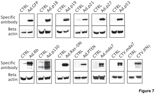
Figure 7. Western blot analysis of Cocca-6A cell lysates following adenoviral transduction.
On the left lane are loaded control Cocca-6A cells. In the lane on right are loaded the transduced Cocca-6A cells. Anti beta-actin was used as a loading control. 50 µg of total lysates were run in SDS polyacrylamide gels. On the left side are indicated the different adenoviral transductions.

