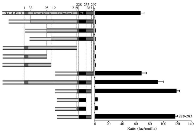
Fig. 2.
Assay of transcription activation by rv-cyclin and rv-cyclin fragments in HeLa cells. Indicated portions of the rv-cyclin protein were fused to the GAL4 DBD, and their ability to activate luciferase expression from the pFR-luc GAL4 reporter construct measured after 48 h. Boundaries of each segment are indicated by a.a. position at the top. The outlines of the predicted cyclin box folds, here designated ‘Cyclin box A’ and ‘Cyclin box B’ are indicated. ‘CC’ represents the putative coiled-coil region. The smallest active region, a.a. 228 – 283, is indicated.

