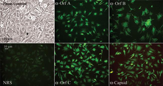
Fig. 1.
Microscopic observations of STECs. Phase-contrast image of confluent STECs illustrates the apical ruffles common in these cells, indicated by an arrow. Immunofluorescent images were labelled with normal rabbit sera (NRS) or with rabbit antisera reactive to the indicated WDSV proteins and fluoresce-inconjugated goat anti-rabbit IgG. Magnification, ×200.

