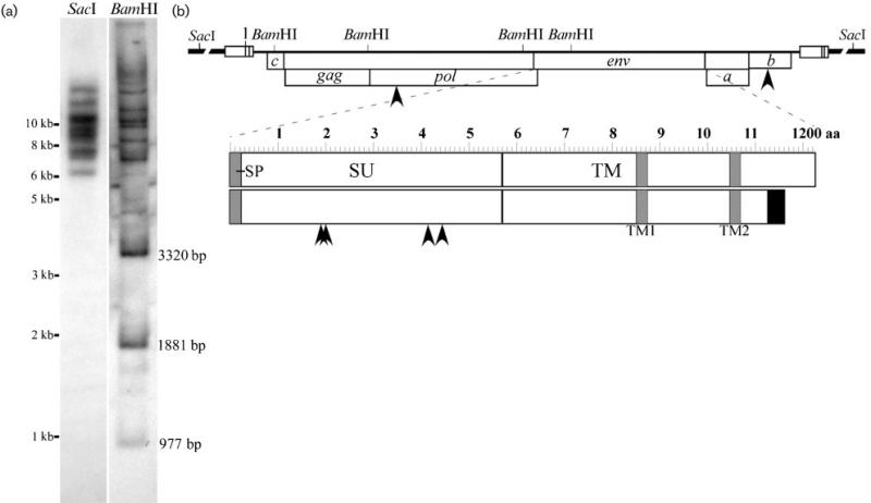
Fig. 2.
(a) Southern blot analysis of STEC chromosomal DNA. SacI liberates whole provirus with adjacent chromosomal DNA. BamHI liberates three internal fragments of fixed sizes as indicated and two external fragments with adjacent host DNA from each provirus. (b) Diagram of the WDSV provirus with adjacent chromosomal DNA (broken line). Open reading frames in frames 1 and 2 are indicated with arrowheads to locate amino acid mutations. The env reading frame is expanded to identify point mutations as well as the revised C terminus with variant sequence (black box) and smaller size. Shaded boxes indicate predicted transmembrane domains, TM1 and TM2, in the transmembrane region (TM), as well as the predicted signal peptide (SP) at the N terminus of the surface region of the protein (SU).

