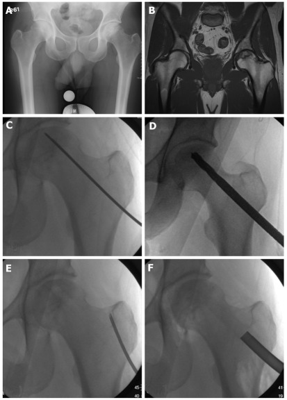
Figure 1.
Forty-one year old male with pre-collapse osteonecrosis of left femoral head as evidenced by (A) plain radiograph and (B) magnetic resonance imaging of pelvis. Patient underwent core decompression: (C) Kirschner wire to localize to affected subchondral bone; D: Drilling of lesion; E: Aspiration of bone marrow from cancellous bone in greater trochanter; F: Insertion of bone graft mixed with bone marrow aspirate.

