
Fig. 1.
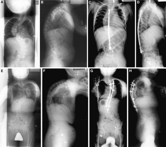
Preoperative anteroposterior (AP) (a) and lateral (b) radiographs of patient 3. Postoperative AP (c) and lateral (d) radiographs after posterior spinal fusion and instrumentation with a single MOE rod. AP (e) and lateral (f) radiographs after the removal of implants due to infection. AP (g) and lateral (h) radiographs postoperatively from final spinal fusion