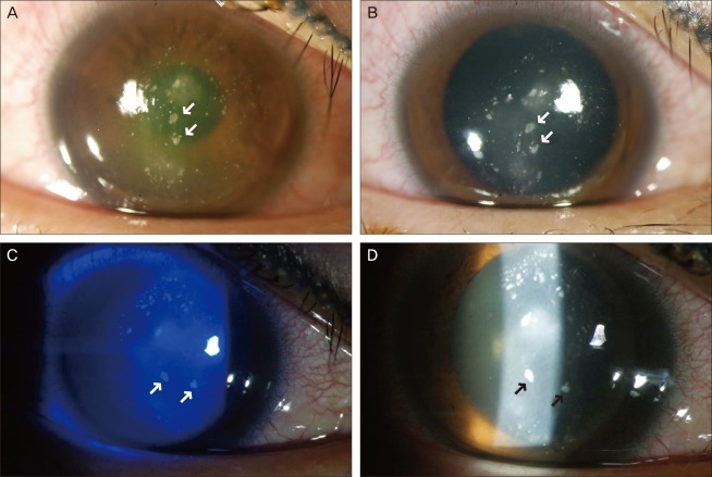
Fig. 1.
Slit-lamp photograph of the patient before flap lifting and irrigation. Upper photographs (A) (before pupillary dilatation) and (B) (after papillary dilatation) show diffusely distributed crystalline materials with stromal infiltration at the laser in situ keratomileusis interface on the first day of hospitalization. Note three large materials distributed linearly in the center of the cornea (see arrows). On the third day of hospitalization, photographs (C) (under cobalt blue filter) and (D) (under slit beam) show the change of distribution of the crystalline materials. Note three large materials in the center that seem to have rotated counterclockwise.

