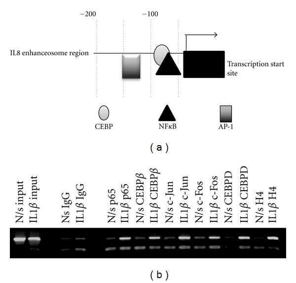
Figure 1.

(a) Schematic of the IL8 promoter enhanceosome region. (b) ChIP analysis demonstrates the in vivo binding of NFκBp65, CEBPβ and d, c-Jun, c-Fos, and H4 to the transcriptional enhanceosome of the IL8 promoter. ChIP assay using antibodies against NFκBp65 and CEBPβ and -d, c-Jun, and c-Fos was performed in nonstimulated (N/s) and IL1B stimulated conditions. The immunoprecipitates were subjected to PCR analysis using primer pairs spanning the IL8 promoter transcriptional enhanceosome. The first two lanes are “input” lanes where no immunoprecipitation was performed prior to PCR. The second two lanes contained IgG antibody for immunoprecipitation (negative controls).
