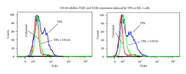
Figure 4.

To determine the role of mitogen-activated protein kinase signal in TPA-induced TLR1 and TLR4 expression in ML-1 cells. Three millions cells were pretreated with 10 μM U0126 for 10 minutes followed by incubation with 5 nM TPA for 24 hours. Cells were then harvested, washed, fixed, permeabilizes, and stained using (Alexa-Fluor-488-) labeled anti-human TLR1 or TLR4 antibodies. The levels of TLR1 or TLR4 protein expression in TPA-treated ML-1 cells were determined by FACS-Scan analysis. Negative controls were stained with isotype-matched- (Alexa-Fluor 488) conjugated IgG and compensation was adjusted using the single-stained cell samples. The fluorescence intensities were determined using Cellquest software (Becton Dickinson, Bedford, MA). Data were analyzed by ModFILT statistical software. The figure depicts representative results from one of three replicate experiments.
