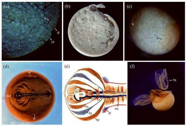
FIGURE 1.
Development of the marsupial frog G. riobambae. (A) Sagittal section of a mid gastrula embryo photographed with differential interference contrast and fluorescence to detect cell borders and Hoechst 33258 stained nuclei. Involuted cells remain in the blastopore lip. The small archenteron (a), dorsal blastopore lip (dl), and yolk plug (yp) are present in the subequatorial region. (B) Sagittal bisection of a late gastrula. The archenteron (a) remains small and cells that involuted during gastrulation form a large circumblastoporal collar (cbc) around the closed blastopore. The blastocoel (b) is still visible. This image was reproduced from BiosciEdNet Digital Library Portal for Teaching and Learning in the Biological Sciences 2010 (http://www.apsarchive.org/resource.cfm?submissionID=3000&BEN=1) (C) The embryonic disk (d) of a late gastrula, stained for cell borders according to del Pino and Elinson.24 The body of the embryo is derived from the embryonic disk. The blastocoel (b) is still detectable. (D) Embryo immunostained for a neural antigen with antibody P3. The embryo is flat, and the heart anlage (ha) develops anterior to the head. On the sides of the embryonic disk, there are preparation artifacts (ar). (E) Composite diagram of neural expression, according to del Pino and Medina.84 The mandibular (m), hyoid (hy), branchial anterior (ba) and branchial posterior (bp) streams of cranial neural crest, neural crest of the trunk (tnc), optic vesicle (ov), midbrain (mb), isthmus (is), rhombomeres (r), neural tube (nt) and pronephros (pr) were detected by expression of antigen 2G9 (brown), ncam protein (dark blue), epha7 (light blue) and pax2 protein (red). Epha7 expression on r3 and r5 is not shown. (F) Advanced embryo immunostained for myosin. In the living condition the disk-shaped bell gills (bg) enveloped the embryo in a vascularized sac.

