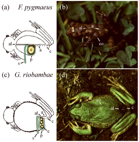
FIGURE 2.

Brooding females of marsupial frogs. (A) Diagram of the pouch and embryos in F. pygmaeus. The anterior limit (al) of the pouch aperture (a) is located behind the head, and the posterior limit is above the cloaca (c). This morphology suggests that the pouch developed from foldings of the dorsal skin during evolution.75 The pouch lining (p) is continuous with the dorsal skin (s). Embryos (e) are brooded inside the pouch. (B) A brooding female of F. pygmaeus. The embryo outlines (eo) are detectable. This small frog, of about 2.5 cm in snout-vent length, carries 6 embryos, each of 3 mm in diameter. (C) Diagram of the pouch and embryos in G. riobambae. The anterior limit (al) of the pouch aperture (a) is located near the cloaca (c). The pouch lining (p) is continuous with the dorsal skin (s) as in F. pygmaeus. Embryos (e) are brooded inside the pouch, which occupies the dorsal and lateral sides of the body in a brooding female. (D) A brooding female of G. riobambae. The embryo outlines (eo) are detectable. The pouch opens above the cloaca (c). This frog measures about 5 cm in snout-vent length and broods about 100 embryos, each of 3 mm in diameter, for about 4 months.65
