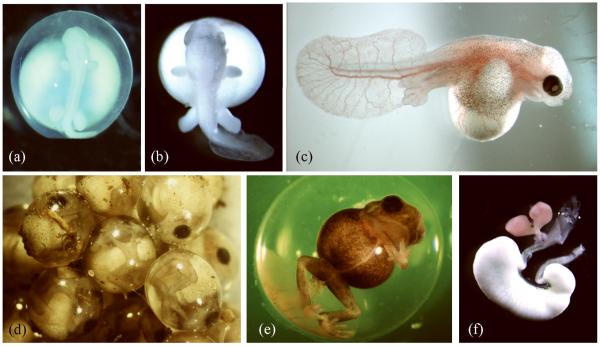
FIGURE 3.
Embryos of the direct developing frog E. coqui. (A) An early E. coqui embryo at Townsend-Stewart (TS) stage 4/5 has developed limb buds and a broad head. (B) By TS7, foot paddles are evident as well as large froglike eyes. (C) This TS10 embryo has been removed from its jelly capsule. The thin, highly vascularized tail serves as a respiratory surface. The pigmented body wall containing somite-derived musculature is extending over the yolk mass to form a secondary coverage. Digits are present and the eye is darkly pigmented. (D) This picture of a clutch of eggs shows TS12 embryos, as they appear naturally in their jelly capsules. (E) A TS14 froglet is about two days from hatching. (F) A digestive tract, dissected from a newly hatched froglet, shows the yolky cells (white) of the nutritional endoderm, attached to the small intestine. Two lobes of liver (pink) and the gall bladder (green) lie between the stomach and the nutritional endoderm.

