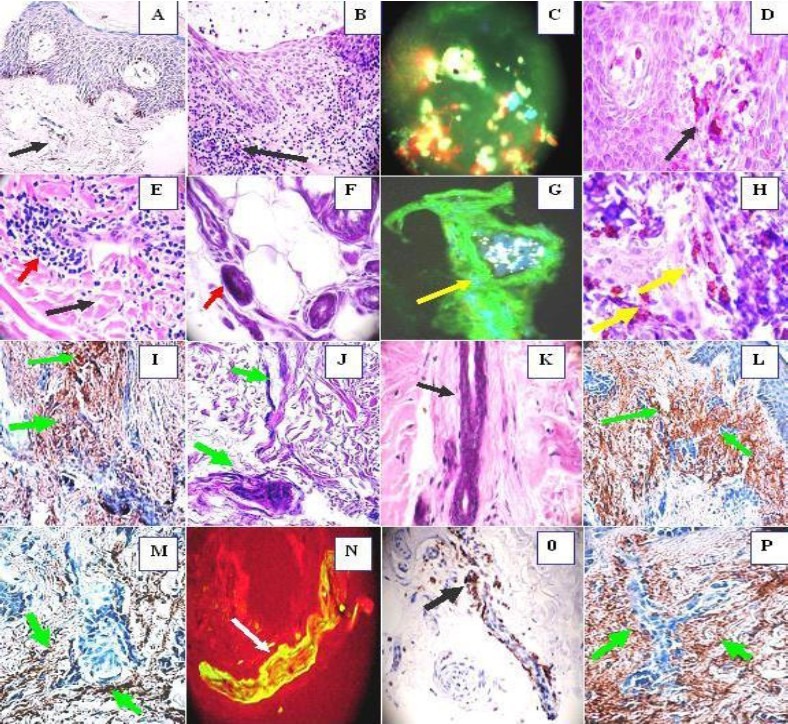
Fig. 2.

IHC (a, d ,h ,i, l, m, o, p), MDIF (c, g, n), H & E (b, e, k). a, Show weak positive stains in the vessels using anti HLA DR, DP, DQ (black arrow). b, Epidermal spongiosis and infiltration of predominantly lymphocytes and histiocytes (black arrow). c. Acrosyringium secretion of IgA (yellow), some nuclei cells debris with Dapi (blue) and positive IgE (green). d. CD8 positive around the superficial vessels (black arrow). e. Sclerodermoid alterations around one sweat glands ductus (black arrow). The red arrows show infiltration of predominantly lymphocytes and histiocytes. f. Necrosis of the sweat glands (red arrow). g. Positive stain of the sweat glands with anti-human fibrinogen FITC conjugated (yellow arrow) (green stain). The white material is self-fluoresce lipofuscin. h, CD8 positive stain around the ecrine ductus under the base membrane zone (yellow arrows). i. Positive stain with anti-human fibrinogen around all the ecrine ductus and sweat glands (brown stain) green arrows. j. Sclerosis of the sweat glands and its ductus (green arrows). K. Narrowing of the sweat glands ductus (black arrow). l. Strong stain (brown) around the sweat glands ductus and its vessels with complement C3C (green arrows). m. Positive stain around the sweat glands coiled portion using complement C3D (brown stain) (green arrows). n. Positive IgE-FITC conjugated in the vessels (yellow stain) (white arrow). o. Positive CD 45 stain along the sweat ductus (black arrow). p. Strong stain (brown) around the sweat glands ductus with complement C3D (green arrows).
