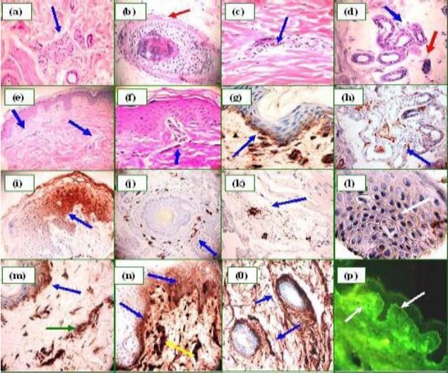
Fig. 1.

H&E sections (a through d). a Displays a mild, perineural, lymphocytic infiltrate (blue arrow) (100X). b. Mild hair follicle spongiosis and a mild, perifollicular, lymphocytic infiltrate (red arrow) (200X). c. Mild dermal perivascular lymphocyte infiltration (blue arrow) (400×). d. Weakly positive PAS staining around the sweat glands (blue arrow); the red arrow indicates a partially necrotic gland (100×). e. Mild dermal edema and perivascular lymphocyte infiltration (e, 100×, and f, 400×) (blue arrows). g. Positive BMZ staining using anti-human fibrinogen (blue arrow). h. The sweat glands display positive staining using anti-human IgM. i, Positive intra-cytoplasmic staining of some keratinocytes. j and k, Positive MCT around some sebaceous glands and/or nearby vessels, respectively. l, Pseudo-pemphigus pattern utilizing C1q (400X). m. Positive BMZ staining (blue arrow) using C3c, and within the superficial vessels (green arrow). n and o. When staining for fibrinogen, positivity was seen at the BMZ and some intracytoplasmic areas of the epidermis (blue arrows), and also around the superficial vessels (yellow arrow). In o, note the anti-fibrinogen positivity around a sweat gland ductus (blue arrows). p Correlating DIF, showing positivity to the BMZ and vessels using anti-fibrinogen, as demonstrated by IHC in o (white arrows).
