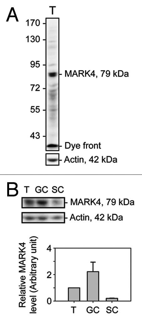
Figure 1. A study to characterize the anti-MARK4 antibody and the cellular distribution of MARK4 in the rat testis. The specificity of the anti-MARK4 antibody was demonstrated by immunoblotting using ~60 μg protein from lysates of adult rat testes using a 6.5% 7 SDS-polyacrylamide gel, with actin as a loading control (A). A prominent band with an electrophoretic mobility of ~79 kDa corresponding to the predicted apparent Mr of MARK4 was detected by immunoblotting. The relative level of MARK4 in lysates from adult rat testes (T), germ cells (GC) isolated from 90-d-old rat testes and cultured in vitro for ~16 h, and Sertoli cells (SC) isolated from 20 d-old rat testes and cultured in vitro for ~4 d were shown in (B), and the composite data were shown in a histogram in (C). Each bar = mean ± SD of n = 3 independent experiments.
