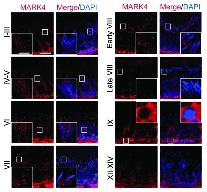
Figure 2. Stage-specific and restrictive temporal and spatial expression of MARK4 in the seminiferous epithelium during the seminiferous epithelial cycle of spermatogenesis. Frozen cross-sections of testes (7 μm in thickness) were obtained with a cryostat at -20°C, fixed in Bouin’s fixative and processed for fluorescence microscopy using an anti-MARK4 antibody (see Table 1).59,71 Cell nuclei were visualized by DAPI (4’,6-diamidino-2-phenylindole) staining. Different stages of the seminiferous epithelial cycle are labeled. MARK4 was detected at the apical ES, BTB and the basement membrane. The expression of MARK4 was found to be stage-specific, with its expression at the BTB being the highest at stages VI-IX. Its expression at the apical ES appeared in stage IX and persisted through early stage VIII, but declined considerably to an almost non-detectable level by late stage VIII. Its localization at the apical ES also changed during the epithelial cycle. For instance, immunoreactive MARK4 was found to surroundthe entire head of elongating spermatids at stages IV-VI, but its localization was shifted and it was found to be restricted to the concave side of the spermatid head at stage VII, and diminished in early stage VIII. In some micrographs, a selected area is boxed and magnified in the inset. Bar in the first micrograph = 50 μm, which applies to all low magnification micrographs; bar = 20 μm in inset, which also applies to all insets.

An official website of the United States government
Here's how you know
Official websites use .gov
A
.gov website belongs to an official
government organization in the United States.
Secure .gov websites use HTTPS
A lock (
) or https:// means you've safely
connected to the .gov website. Share sensitive
information only on official, secure websites.
