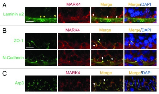
Figure 4. A study by dual-labeled immunofluorescence analysis to illustrate the localization of MARK4 at the hemidesmosome/basement membrane, the BTB, and the apical ES. Precise localization of MARK4 in the seminiferous epithelium was further investigated by its co-localization with corresponding markers of the hemidesmosome/basement membrane (e.g., laminin α2) (A), the BTB (e.g., ZO-1, a TJ adaptor; N-cadherin, a basal ES protein) (B), and the apical ES (e.g., Arp3)(C). The general site where hemidesmosomes are found is annotated by “yellow” arrowheads, and selected merged signals are annotated by “white” arrowheads. Bar in the first micrograph in A, B or C = 40 μm, which applies to remaining micrographs.

An official website of the United States government
Here's how you know
Official websites use .gov
A
.gov website belongs to an official
government organization in the United States.
Secure .gov websites use HTTPS
A lock (
) or https:// means you've safely
connected to the .gov website. Share sensitive
information only on official, secure websites.
