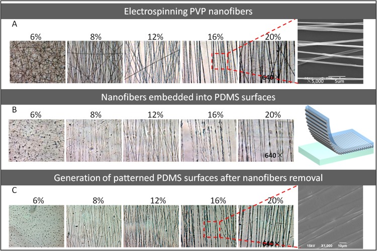
Figure 2.
The photographs of the electrospun PVP nanofibers and produced patterned PDMS surfaces under different concentrations. (a) The microscope images of the PVP nanofibers spun with five concentrations (640×). SEM image of PVP nanofibers spun with 16% initial concentration. (b) The microscope images of PDMS surfaces embedded with PVP nanofibers (640×). (c) The microscope images of patterned PDMS surfaces after fibers removal (640×). SEM image of PDMS surfaces patterned with microstructures.

