
Figure 1.
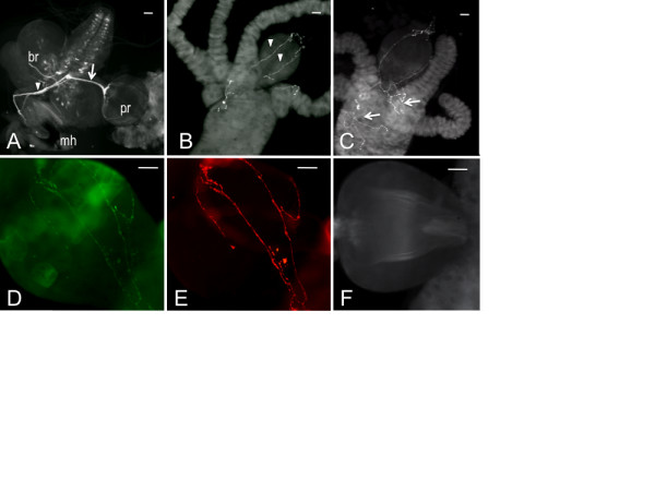
The Drosophila larval feeding circuit is not innervated by dopaminergic fibers. Gut tissues dissected from Drosophila 3rd instar larvae were immunostained with an antibody raised against Drosophila neuronal tryptophan hydroxylase (DTRH, A) or 5-HT (B, C). A. mh, mouth hooks; pr, proventriculus; br, brain (showing the pattern of 5-HT neurons). The arrowhead designates the frontal nerve and an arrow the recurrens nerve. B. proventriculus showing axonal fibers (denoted with arrowheads). C. fibers fasiculating in the midgut (denoted with arrows). D - E. proventricular fibers from larvae carrying a neuronally expressed green fluorescent protein tagged to synaptotagmin. E, GFP fluorescence; D, anti-DTRH. F. Gut tissues stained with an antibody raised against Drosophila tyrosine hydroxylase (DTH). Scale bars = 20 μm.