Figure 5.

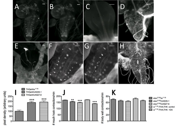
Constitutive induction in neuronal DTH or increased levels of systemic DA during late embryogenesis results in depressed feeding behavior in larvae. Two independent UAS transgenic lines (35.1 and 27.6, on chromosomes 2 and 3, respectively) were used to increase neuronal DTH levels. A - C. DTH immunoreactivity in the ventral ganglion of w1118/UAS27.6 larvae. A. Dorsolateral neurons. B. Medial neurons). C. Proventriculus. D - E. DTH immunoreactivity from elavC155/UAS27.6 larvae. D, larval brain. E. Proventriculus visualized with anti-DTH. F - H. DTH immunoreactivity in sibling progeny from a UAS35.1/Bc1 x elavC155 cross (Bc [Black cell] is a larval marker). F. Dorsolateral DA neurons visualized with anti-DTH in a Bc1/UAS35.1 larval ventral ganglion. G. Medial DA neurons visualized with anti-DTH in a Bc1/UAS35.1 3rd instar larval ventral ganglion. H. DTH immunoreactivity in an elav/UAS35.1 larval brain. Scale bar = 50 μm. I. Relative pixel intensity of the most distal pair of dorsolateral neurons in THGal4/w1118, THGal4/UAS35.1 and THGal4/UAS27.6 larvae. Third instar larval brains from the different genotypes were dissected and incubated with anti-DTH antibody. Brains from all animals were assessed in parallel. THGal4/w1118, n = 12 neurons [control]; THGal4/UAS35.1, n = 24 neurons; THGal4/UAS27.6, n = 15 neurons. ***p < 0.001, one-way ANOVA followed by Tukey's multiple comparisons post-test. The animals were then assayed for feeding (J) and compared with 16 hr old control (w1118;THA;THK) embryos were exposed to serum-free media with or without 10-6M DA for 6 hours until hatching. K. Locomotion was unaffected. **p < 0.01, ***p < 0.001, one-way ANOVA followed by Tukey's multiple comparisons post-test. n = 40 for behavioral analyses, from 4-6 independent experiments. Lines above the graph depict standard error of the mean.
