
Figure 1.
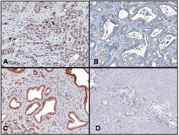
Staining for RRM1 and ERCC1 proteins. (A) ERCC1-positive sample. Note the intense nuclear staining. (B) ERCC1-negative sample. (C) RRM1-positive sample. Note the intense cytoplasmic staining. (D) RRM1-negative sample.
Official websites use .gov
A
.gov website belongs to an official
government organization in the United States.
Secure .gov websites use HTTPS
A lock (
) or https:// means you've safely
connected to the .gov website. Share sensitive
information only on official, secure websites.
Staining for RRM1 and ERCC1 proteins. (A) ERCC1-positive sample. Note the intense nuclear staining. (B) ERCC1-negative sample. (C) RRM1-positive sample. Note the intense cytoplasmic staining. (D) RRM1-negative sample.