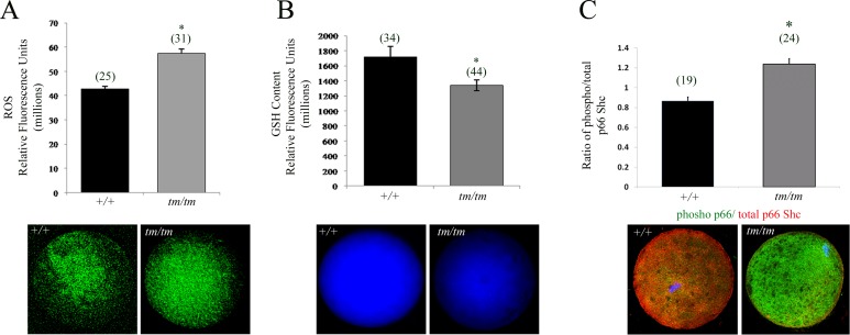
FIG. 2.
Altered GSH levels result in increased ROS production in Nlrp5-deficient oocytes. A) ROS measurement in Nlrp5+/+ and Nlrp5tm/tm oocytes. Values represent relative fluorescence units ± SEM. Increased ROS production was observed in Nlrp5 hypomorph oocytes. Asterisk (*) indicates significance; P < 0.05. Representative images are shown below. B) Baseline GSH content in Nlrp5+/+ and Nlrp5tm/tm oocytes. Values represent mean fluorescence units ± SEM. A significant reduction in the baseline GSH content was observed in tm/tm oocytes (*P < 0.05). C) Expression of p66 SHC in oocytes. Relative fluorescent intensity in RFU generated by phospho p66 (green) to total p66Shc (red) was used to show increased expression of phosphorylated isoform (P < 0.001). There was no change in the signal intensity of total p66SHC, and change in the ratio is due to rise of phospho p66SHC signal (green). Numbers in parentheses represent number of oocytes used in each experiment. Original magnification ×200.

