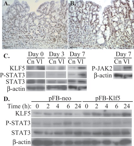
Figure 4. Klf5 overexpression in intestinal epithelia increases STAT3 phosphorylation in vivo.
(A, B) Immunohistochemistry of colonic mucosa from control (A) and Villin-Klf5 (B) mice following DSS treatment revealed more phosphorylated STAT3 in regenerating colonic epithelial cells from Villin-Klf5 mice. (C) Western blot of colonic epithelial scrapings from control (Cn) and Villin-Klf5 (Vl) mice at the indicated time points following DSS treatment revealed increased phospho-STAT3 and phospho-JAK2 in Villin-Klf5 mice at day 7, while phospho-STAT3 was decreased in Villin-Klf5 mice at day 0 and unchanged at day 3. (D) Western blots confirmed increased KLF5 expression in IEC-6 cells transduced with pFB-Klf5 compared to pFB-neo. However, following linear wounding, no induction of phosphorylated STAT3 was observed in pFB-Klf5 infected cells, compared to control pFB-neo infected cells.

