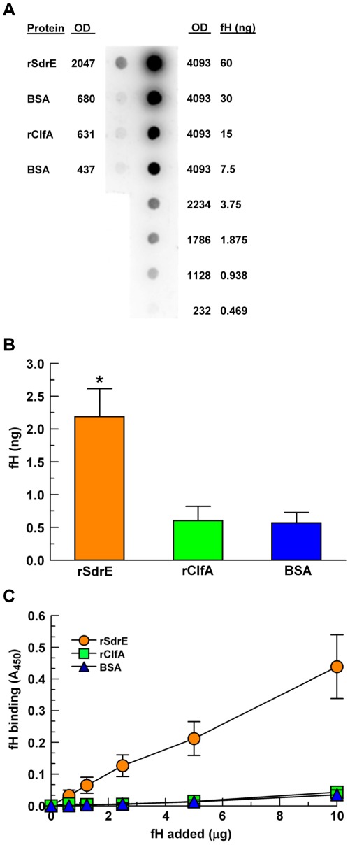
Figure 3. Purified fH binding to recombinant proteins.
A, A representative fH overlay dot blot using a purified fH dilution series as a quantitation control (right column, fH). rSdrE, rClfA and BSA (5 µg) were adsorbed to a PDVF membrane (left column), blocked, then overlaid with purified fH (20 µg in 10 ml block buffer); BSA was used as a control. fH binding was determined via optical densitometry using the purified fH dilution series as a standard curve. B, Quantitative fH binding via dot blot, as described in (A), *p<0.01; n = 4. C, Modified ELISA: rSdrE, rClfA and BSA were adsorbed to a microtiter plate and incubated with various amounts of purified fH for 1 hr; data represent the mean of at least three independent experiments ± SEM.

