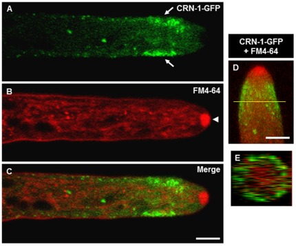
Figure 1. Subapical localization of coronin.
(A) CRN-1-GFP forms a subapical collar along the inner perimeter of the hypha (arrows), (B) FM4-64 staining reveals the position of the Spk (arrowheads), (C) merge of CRN-1-GFP and FM4-64 staining shows the absence of CRN-1-GFP in the Spk, single confocal plane images. (D) 3D reconstruction of merged confocal z-stacks showing CRN-1-GFP and FM4-64 localization, (E) orthogonal view of the 3D reconstruction shown in (D), the yellow line indicates the position within the tip where the cross-section was taken. Scale bars = 5 µm.

