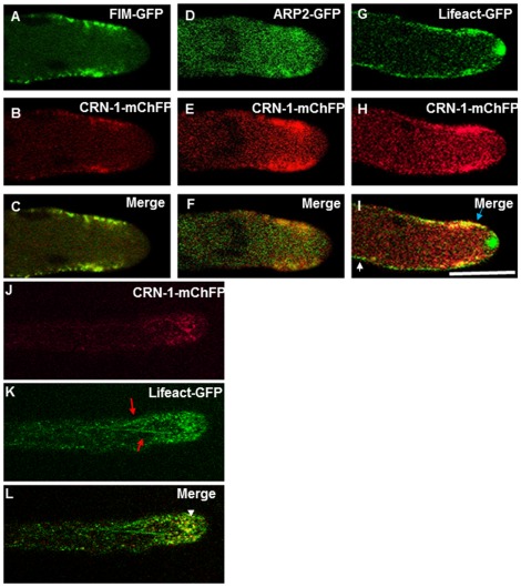
Figure 2. Co-expression of coronin with fimbrin, Arp2 and actin.
(A–C) Colocalization of Fimbrin (FIM-GFP) and CRN-1-mChFP. (D–F) Colocalization of Arp2 (ARP-2-GFP) and CRN-1-mChFP. (G–I) Partial colocalization of the actin marker Lifeact-GFP and CRN-1-mChFP. (J–L) Co-expression of CRN-1-mChFP and Lifeact-GFP showing the lack of colocalization between coronin patches and actin cables. are depicted by. (L) Merge, not clear association of crn-1 patches is observed with actin filaments, arrowhead shows colocalization of actin patches with CRN-1-mChFP. The white arrow points a region where there is only labeling with Lifeact-GFP and the blue arrow show the patches where CRN-1-mChFP and Lifeact-GFP colocalized. Note the presence of actin in the Spk but not of patch related ABPs. The red arrows in (K) point the actin cables and the white arrowhead show the colocalization of actin and coronin in the patches subapical collar. Scale bar = 5 µm.

