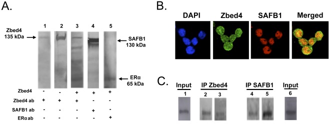
Figure 7. Zbed4 interacts with SAFB1 in vitro and in vivo.
A. Membrane overlay assay: Proteins were resolved on SDS-PAGE and transferred to a PVDF membrane. Lane 1: cell lysate; lane 2: purified Zbed4 protein; lanes 3, 4 and 5: cell lysate incubated with Zbed4 protein (overlay). Lanes 1, 2 and 3 were probed with the Zbed4 antibody and lanes 4 and 5 with the SAFB1 and ERα antibodies, respectively. B. Subcellular co-localization of Zbed4 with SAFB1, carried out as described in Materials and Methods. The merged image clearly shows that Zbed4 co-localizes with SAFB1 in the nucleus of Y79 retinoblastoma cells. DAPI was used to stain the nuclei. C. Co-immunoprecipitation experiments were performed using Y79 retinoblastoma cell extracts. Both Zbed4 and SAFB1 were detected in the immunoprecipitated proteins obtained with Zbed4 antibody (left panel) or SAFB1 antibody (right panelIn each case, each duplicate lane of the blots obtained after SDS-PAGE of the immunoprecipitated proteins was incubated with Zbed4 or SAFB1 antibodies. Immunoprecipitation experiments were performed using Y79 retinoblastoma cell extracts (Input) and antibodies against Zbed4 or SAFB1. Duplicate aliquots of the proteins immunoprecipitated by each antibody were immunoblotted after SDS-PAGE with antibodies against Zbed4 (lanes 2 and 4) or SAFB1 (lanes 3 and 5). Both Zbed4 and SAFB1 are detected in each immunoprecipitate. Aliquots of Input material also show on the Western blots the presence of Zbed4 (lane 1) and SAFB1 (lane 6).

