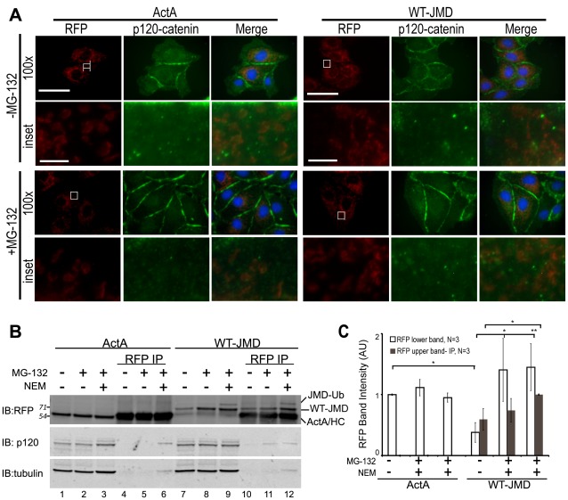
Figure 5. Localization and binding of WT-JMD and p120-catenin.
(A) Immunofluorescence of MDCK cells transiently expressing ActA or WT-JMD. Images for RFP (red), p120-catenin (green) and merged are shown separately (100×). Boxed areas are shown as higher magnifications below (RFP-, p120- and merge-inset). All images were from the same experiment and processed identically between cell lines. Scale bar is 25 µm in 100× images, and 5 µm in insets. (B) Lysates and RFP immunoprecipitates (IP) of ActA and WT-JMD stable cell lines under normal conditions, upon proteasome inhibition, or NEM treatment (to inhibit de-ubiquitinating enzymes). Immunoblots (IB) for RFP show: a slower migrating band (upper band-JMD-Ub) that appears only in WT-JMD cells in the presence of MG-132 and NEM; the band identified as ActA/HC comprises a co-migrating ActA and the IgG heavy chain (HC). Number(s) on the side of the gels are the apparent molecular weights of protein standards (× 10∧3). (C) Quantification of RFP intensities normalized to tubulin in WT-JMD stables cell lines. Data averaged from 3 independent experiments (+/− s.e.m.), and 2 independently cloned stable cell lines; *p≤0.05, **p≤0.01.

