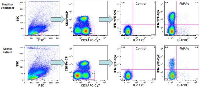
Figure 1. Strategy for the analysis of Th1 and Th17 lymphocytes.
Dot plots shown are representative of one healthy volunteer and one septic patient. T cells were identified based on CD3-APC-Cy7 staining and forward scatter versus side scatter parameters. For the analyses, T helper cells were gated as CD3+CD8−, and the percentages of cells producing IFN-γ or IL-17 were determined with the quadrants established based on the control samples.

