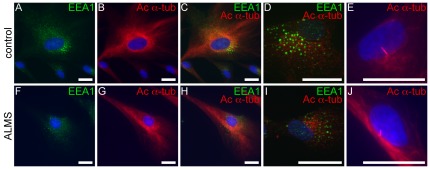
Figure 5. Immunostaining of microtubules and endosomes in ALMS fibroblasts.
(A–D, F–I) Patient and control fibroblasts are stained with EEA1 (green, early endosomal marker) and acetylated α-tubulin (red, microtubule markers). Panels E & J show formation of primary cilia in differentiated fibroblasts from patients and controls. Scale bars = 25 µm.

