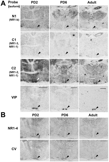
Figure 4. NR1 variable region gene expression in the Siberian hamster forebrain at different developmental ages.
A. Autoradiographs of adjacent coronal sections from PD2 (left), PD6 (middle) and adult (right) hamster brain hybridized with 35S-labelled oligonucleotide probes N1, C1 and C2 complimentary to NR1 variable regions. Dense hybridization in the SCN only occurs with the C2 probe. Hybridization of the VIP probe identifies the position of the ventro-lateral SCN (bottom). Age-specific sections are from same representative animal. Arrows indicate position of SCN. Scale bars = 1000 µm. B. Hybridization of the NR1-4 probe (top), and adjacent cresyl violet stained section (bottom) in the hamster SCN. Autoradiographs of coronal sections taken through the forebrain at the level of the SCN (arrow) at the ages PD2 (left), PD6 (middle) and adult (right). Age-specific sections are from same representative animal. Cresyl violet Nissl stain (CV) identifies the SCN region by its dense regional staining. Scale bars = 1000 µm.

