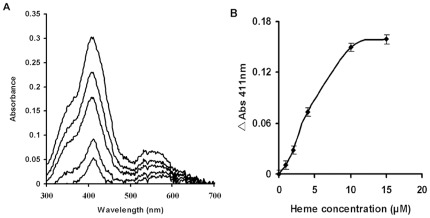
Figure 4. Binding of heme to HemS.
(A): Increasing amounts of heme (1 µM–20 µM final concentration) were added to HemS (10 µM) as decribed in “Materials and Methods” and the spectrum (300 nm–700 nm) was recorded after 5 min for each addition. The Soret band at 411 nm increases with each addition of heme as demonstrated by absorbance peak increases at 411 nm. (B): Absorbance at 411 nm was measured for each sample and plotted versus heme concentration. Experiments were performed in triplicate and a single representative experiment is presented.

